
Cardiac vasculature
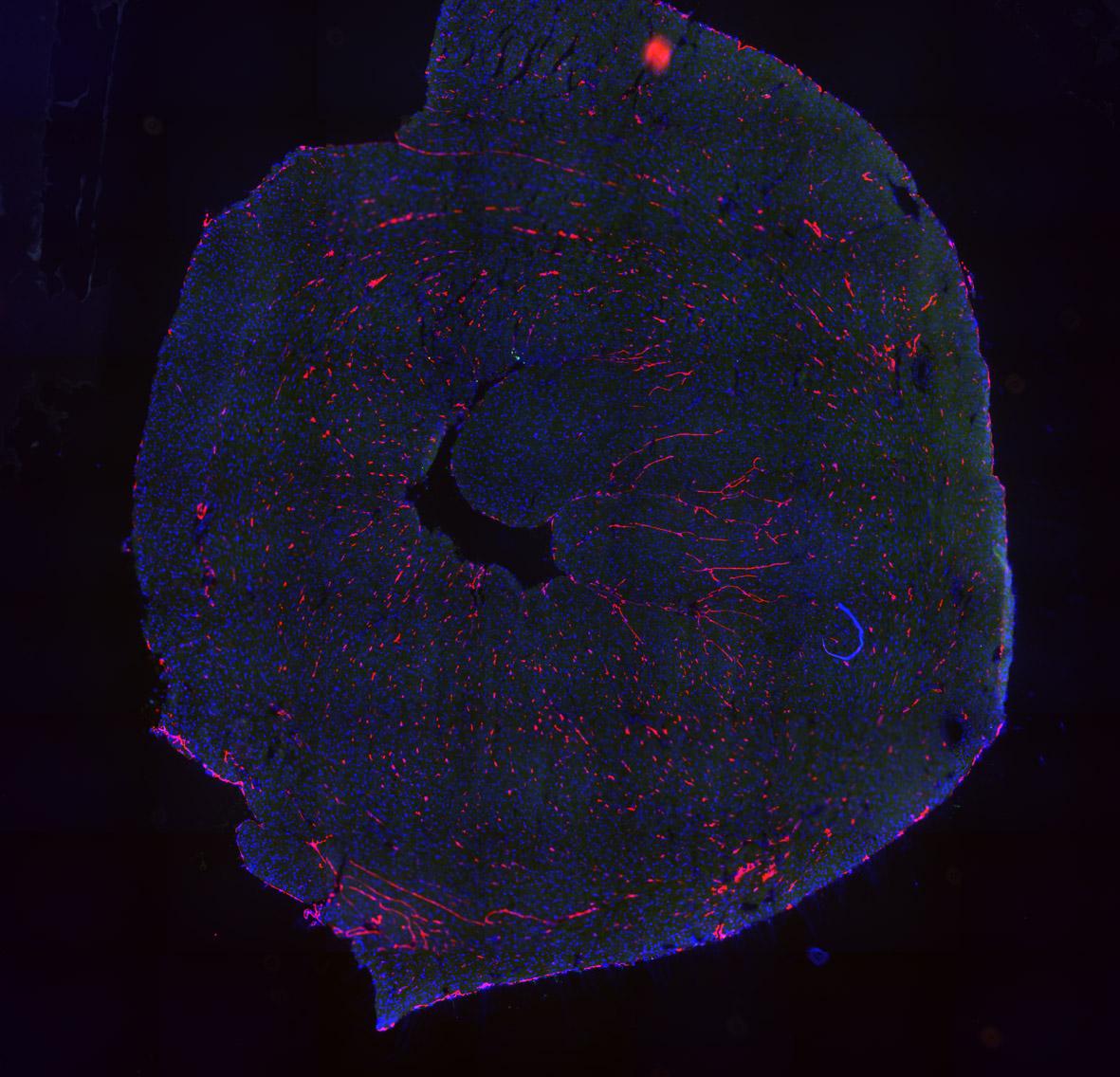
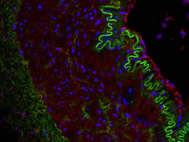
We employ different imaging techniques to visualize cardiac blood vessel: macroconfocal imaging, traditional immunohistochemistry of vascular endothelial or arterial markers in cardiac sections, and confocal or light sheet imaging of whole-mount sections
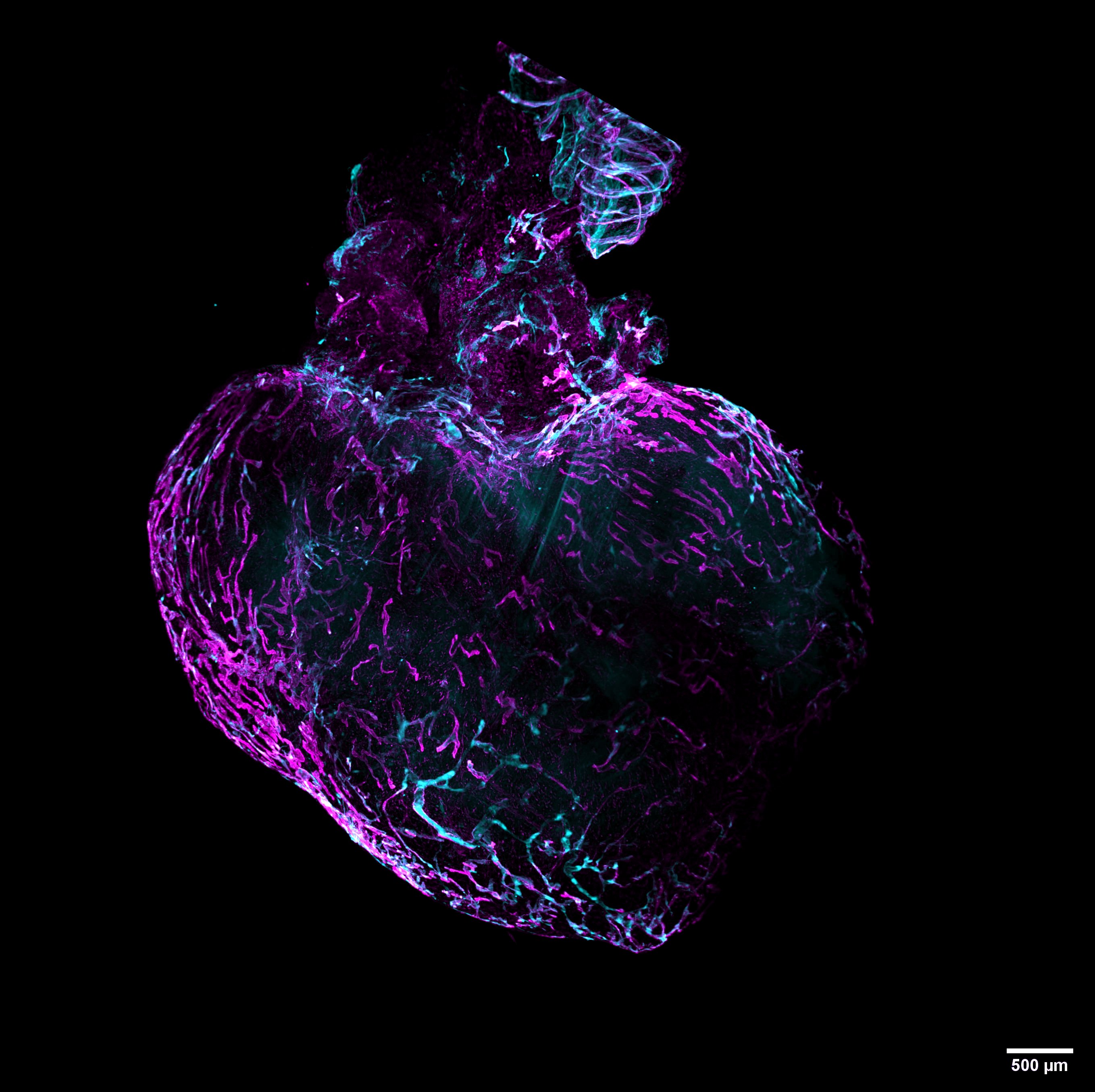
Coraline Heron, a PhD student in the laboratory, has together with our collaborator, David Godefroy (Inserm U1239), used light sheet imaging to reveal the delicate network of cardiac lymphatics in mice. Their work was awarded with a 1st Prize at the yearly FASEB BioArts competition in 2020.